|
|
Post by aqt on Mar 25, 2010 17:20:38 GMT -5
en.wikipedia.org/wiki/NanobacteriumNanobacteria (singular nanobacterium) is the name of a proposed class of living organisms; specifically cell-walled microorganisms with a size much smaller than the generally accepted lower limit size for life (about 200 nanometres for bacteria). Originally based on observed nano-scale structures in geological formations (including some meteorites), the status of nanobacteria is controversial, with some researchers suggesting they are a new class of living organisms[1][2] capable of incorporating radiolabeled uridine,[3] and others attributing to them a simpler, abiotic nature.[4][5] One skeptic dubbed them "the cold fusion of microbiology", in reference to a notorious episode of erroneous science.[6]The term 'calcifying nanoparticles' (CNPs) has also been used as a conservative name regarding their possible status as a life form. The most recent research tends to agree that these structures exist, and probably replicate in some way.[7] Their status as living entities is still hotly debated, though some researchers now claim that the case that they are nonliving crystalline particles is conclusively proven.[8] In medicine, they have been implicated in the formation of both kidney stones and arterial plaque.
|
|
|
|
Post by aqt on Mar 25, 2010 17:23:52 GMT -5
nanotech.biz/i.php?id=2002_01_25Question 1: Tell us about how and when you discovered nanobacteria. Our colleagues and Nobel Prize Nominee Medical Researchers, Neva Ciftcioglu, PhD and Olavi Kajander, MD, PhD, (Microbiology Department-University of Kuopio, Finland) were working on several medical research projects in 1992 using mammalian cell cultures. They became frustrated because their mammalian cell cultures kept dying. Mammalian cell cultures and most all human biologicals are normally grown in fetal bovine serum (FBS) and cell culture death is a common problem in medical research that forces researchers to start entire projects over again and again. Well, this time instead of just discarding the dead mammalian cell cultures, they just left them in the incubator and subsequently forgot about them. Approximately four months later finding the old cultures, they realized that an unusual thin slimy film had developed on the culture surfaces. Well, since scientists are by nature as inquisitive as cats, Drs. Ciftcioglu and Kajander they couldn’t resist full evaluation of this unusual slime. They were not able to determine the nature of the film using the highest power light microscopes, so they used Scanning Electron Microscopes and Transmission Electron Microscopes from lowest to highest magnification (100,000X). Lo and behold! They found these 20-200nanometer sized nanobacteria in calcified shells: The bacteria in the stone! They had never seen anything like them, and nothing like them had ever been seen or described in the microbiology world. These nanobacteria are structurally and physiologically unique in many ways: they are 20-200 nanometers in size, they have a unique “cellular” structure and membrane structure like nothing else on earth, they replicate very slowly (every 3-5 days) and by different methods, they are pleomorphic meaning that they assume different life forms for different phases and activities of their lives, they can go dormant in a self-made calcium shell, they are saprophytic in humans, they are “the toughest of bugs” resistant to being killed both In-Vitro and In-Vivo and they are the cause of many human diseases. They are structurally and functionally simplistic, unbelievably small and genetically unique. Although nicked-named by one reporter as “Conan the Bacterium”, Drs. Ciftcioglu & Kajander formally named this novel pleomorphic nanobacteria that thrives in our blood, Nanobacterium sanguineum.
|
|
|
|
Post by aqt on Mar 25, 2010 17:27:18 GMT -5
tiny.cc/k52x7Quote: Katinka "Nanobacteria.. biofilm producing....protectiv shell...feeding on heavy metals...Ascorbic acid and Salt....Detoxification. This all sounds familiar to me. Marc mentioned Biofilms with hardening shells on his site. Could be that this is the reason why the VitaminC/Salt protocol works against Morgellons..(for me it did) together with detox and immune system upbuilding. The symptoms of Nanobacterium sanguineum are similar to Morgellons Disease. NanobacLabs also has the NanobacTEST-U/A, a rapid urine screening test for nanobacteria that can be done in a doctor's office with results available in minutes. "
|
|
|
|
Post by aqt on Mar 25, 2010 17:31:20 GMT -5
Morphological, cultural, and immuno-histochemical characteristics of “Nanobacterium sanguineum” (NB) described in the literature are reviewed. NB is reported to be a motile, Gram negative organism that divides by binary fission within a calcium-coated slimy shell; this yeast-like shell replicates by budding. It measures between 20 and 200nm with a unique structure containing 16S ribosomal RNA. NB has been observed by electron microscopy in coronary artery plaques (CAD) and in kidney stones (KS) found in renal diseases. On the basis of supportive literature, we suggest that NB is not only present in the human body but also has auxiliary association with human ailments without a specific etiological role; anti-NB antibody has been detected in subjects with calcified lesions and inflammation in diverse ailments including choriodecidual inflammation in pregnancy, ovarian cancers, arthritis and even Alzheimer’s disease. More recent report on the detection and vertical transmission of NB antigen and anti-NB antibody in HIV-infected mothers supports the view that NB might be an important opportunistic infective agent contributing to HIV pathology; we note that the presence of viable and transmitting NB was not studied and suggest further studies to establish vertical transmission of NB in HIV-infected persons. On the basis of the foregoing we suggest that NB possibly exacerbates human ailments and raise the question: Is NB a new life-form in search of human ailment or a commensal organism? Recognizing the presence of NB in the human body, we discuss clinical trials, reported in the literature relevant to its eradication, with a rectal suppository containing very high amounts of disodium EDTA and tetracycline. We suggest that tetracycline in this formulation acted in combination with EDTA, more as a chelating agent than an antibiotic; oxytetracycline- a non-chelating form of tetracycline-does not inhibit or kill NB. Evaluation of anti-NB effect of orally administrable and potentially safer as well as therapeutically more acceptable chelating agent - ascorbic acid, acting alone or in combination with antibiotics-that eradicates another slime forming bacterium – Pseudomonas aeruginosa – in children with cystic fibrosis, is suggested.tiny.cc/k52x7
|
|
|
|
Post by aqt on Mar 25, 2010 17:33:10 GMT -5
|
|
|
|
Post by aqt on Mar 25, 2010 17:36:03 GMT -5
Similarly, since 1990 there has been mounting evidence that abnormal calcification may be caused by a bacterial organism about 1/100 the size of a conventional bacterium, called Nanobacterium sanguineum (“nano” is the Latin word for very small or minute). While other microbes like Chlamydia and assorted fungi have been implicated in the development of abnormal calcification, research indicates that the more likely cause may be nanobacteria. www.alive.com/3140a6a2.php?subject_bread_cramb=218aqt |
|
|
|
Post by aqt on Mar 25, 2010 17:39:49 GMT -5
|
|
|
|
Post by katinka on Mar 25, 2010 18:23:50 GMT -5
uh..and..obviously you've found mine too!..  ...I recognized my typical way of writing.. I've done quite a bit research on Nanobacteria a while back also on LB. Should I add it here? Kat |
|
|
|
Post by skyship on Mar 25, 2010 23:11:06 GMT -5
there has been some new nano bacteria that involves the archaea in it.
that archaea I believe is involved in the kinesin. maybe the submicron particle
that starts the nano growing. cross between organic/inorganic
live cell and rocks. to be blunt.
I would appreciate if you have studied any of the links between live bacteria, microbes,
and inorganic metals and minerals, the biomineralization.
thank you and I would love to read more on this.
skyship
|
|
|
|
Post by skyship on Mar 25, 2010 23:21:42 GMT -5
HEre is a description of spines, and flagella etc. "Most commonly found on the surface of bacteria are flagella used for swimming (47); the type III secretion injectisome (needle structure) (21), which is used to deliver effector molecules from pathogenic organisms into host cells; and a wide variety of thinner organelles that fall under the broad designation of pili (13, 33, 58, 64, 69, 78). Different classes of these structures (type I pili, type IV pili, sex pili, etc.) which differ significantly in their structure, assembly, and function have been identified. Their many roles include adhesion, twitching (or surface) motility, and delivery of DNA and toxins, as well as functioning as electrically conductive "nanowires." Other, less commonly studied appendages have also been reported, such as spinae (9). Archaea, representing the third domain of life, have been isolated from the most extreme environments known to harbor life (15). They have been cultivated from environments that mark the upper limits of life in regard to temperature, pH, and salt concentration. Furthermore, molecular techniques have indicated that archaea are common in a variety of less extreme niches, such as ocean waters, freshwater sediments, and soil (20, 23, 66). Evidence points to a broader and more significant role in the ecosystem for archaea than previously believed (12, 14, 49).".......... ......"Some, like the archaeal flagella and pili, appear at first glance to be similar to their bacterial counterparts but possess significant differences and archaeal twists. Other structures, such as cannulae and hami, appear to be unique to the archaeal domain. Still other structures, such as the bindosome, are predicted to exist from genetic and biochemical data although direct electron microscopic evidence is, as yet, lacking. Studies on archaeal extracellular structures have enriched our understanding of a variety of topics, including protein export, posttranslational modifications, assembly mechanisms, and metabolism. They have also provided unique examples of adaptations to their environments not observed in bacteria. The capacity of some archaea to assemble these structures in extreme conditions that bacterial structures often fail to withstand is of great interest from both fundamental and applied viewpoints.".. jb.asm.org/cgi/content/full/190/18/6039Photo of Pyrodictium cell with cannulae with tubules. The matrix......... jb.asm.org/cgi/content/full/190/18/6039/F4--------------------------------------- lil sissy, The cannulae, forms on pancreas, they called it cannicula, has to be the mineral stone deposits in pancreas, like kidney and gall stones. I think the nanobacteria forms from the archaea and human gene or fungi, possibly the Kammy GP fungi, or arbus. she found. Has the sphere in it. What do you think? skyship |
|
|
|
Post by skyship on Mar 25, 2010 23:26:06 GMT -5
Kammy, Could your carbon ball be the Hami? jb.asm.org/cgi/content/full/190/18/6039/F5check this out. It has a coccus and would be the eukaryote. Mr. Carnicom says it is a link between these three kingdoms. eukaryote, archaea, and bacteria. this is very close. ================== says here hami is initiation of the biofilm: Recently, in a sulfidic spring near Regensburg, Germany, slime-like, milky drops consisting almost entirely of the SM1 euryarchaeon were harvested (35). This organization differs from the previously described string-of-pearls structures in several remarkable ways. While the archaeon-to-bacterium ratio in the string-of-pearls community is approximately 1:1, the SM1 biofilm is dominated by archaeal cells, representing the first archaeal monospecies biofilm in nature. Confocal laser scanning microscopy revealed a constant and regular three-dimensional arrangement of the archaeal cells, with cells being approximately 4 µm apart. The regular distance is speculated to be caused by the hami of neighboring cells, which have an average length of 2 µm. The hamus also forms the main protein component of the extracellular polymeric substances, thereby contributing to the biofilm structure. Several gaps, resembling the typical water channels in bacterial biofilms, were also observed. Henneberger and colleagues (35) proposed that the hami function in surface attachment and biofilm initiation, much like flagella and pili can in bacterial biofilm formation." jb.asm.org/cgi/content/full/190/18/6039skyship skyship |
|
|
|
Post by skyship on Mar 25, 2010 23:31:08 GMT -5
aqt,
thank you for reposting this thread, sent me a new direction.
I do believe that archaea is the submicron particle,
what do you think?
the cross into the inorganic, could be from moon or mars or volcano or under sea.
but, at one time came from "out there"?
what do you think?
skyship
|
|
|
|
Post by skyship on Mar 25, 2010 23:38:57 GMT -5
Photo of pili jb.asm.org/cgi/content/full/190/18/6039/F6precursor protein or BINDOSOME Model of the assembly of surface structures in the cell envelope of Sulfolobus solfataricus: Precursor proteins (SBPs, prepilins, or preflagellins) are processed by PibD and are then inserted by their specific assembly system either in the bindosome structure, the UV inducible pili or the flagellum. The exact nature of the bindosome structure is not known yet, and an alternate format, shown attached to the S-layer, is also indicated. All three assembly systems share the same core of the machinery: an integral membrane protein and a cytoplasmic ATPase. (B) Electron micrograph of flagellated S. solfataricus P2 cells. Flagella are present all around the cells and do not appear bundled. Bar, 1 µm. jb.asm.org/cgi/content/full/190/18/6039/F7Bindosome: An interesting proposed archaeal structure of unique function is the bindosome in S. solfataricus (2). So far, the actual structure has not been visualized in the membranes of S. solfataricus, but rather it is thought to be a pilus-like structure close to the cytoplasmic membrane or integrated within the S-layer (Fig. 7). The main evidence in support of the presence of this hypothesized structure is that the proposed structural components, the substrate binding proteins (SBPs), contain class III signal peptide sequences, a feature typical of proteins which are well known to form oligomeric structures in both archaea and bacteria. The oligomerized complex is proposed to play a role in facilitating sugar uptake, a function that enables S. solfataricus to grow on a broad variety of substrates. Sulfolobus species are hyperthermophilic acidophiles typically found in volcanic springs, with optimal growth at around pH 2 to 3 in the temperature range of 75 to 80°C. One interesting distinction that draws S. solfataricus apart from other Sulfolobus species, such as S. tokodaii and S. acidocaldarius, is the ability to grow on a wide variety of sugars as its only carbon source. Previous studies showed that S. solfataricus has a wide range of ABC transport systems for sugar uptake (4, 26). ABC transporters encompass the actual transport domain in the membrane and cytoplasmically located ATPases, which drive the transport of the substrate by ATP hydrolysis from : Cell Surface Structures of Archaea{triangledown} jb.asm.org/cgi/content/full/190/18/6039#F7skyship |
|
|
|
Post by katinka on Mar 26, 2010 4:29:35 GMT -5
|
|
|
|
Post by katinka on Mar 26, 2010 5:57:46 GMT -5
Nanoarchaeum equitansen.wikipedia.org/wiki/Nanoarchaeum_equitansSince it grows in temperatures approaching boiling, it is considered to be a thermophile. Nanoarchaeum appears to be an obligatory symbiont on the archaeon Ignicoccus; it must be in contact with the host organism to survive. Nanoarchaeum is peculiar in that its 16S RNA sequence is undetectable by the most common methods. Its cells are only 400 nm in diameter, making it the next smallest known living organism, excepting possibly nanobacteria and nanobes. Nanoarchaeum cannot synthesize most nucleotides, amino acids, lipids, and cofactors. The cell most likely obtains these biomolecules from Ignicoccus. However, unlike many parasitic microbes, Nanoarchaeum has many DNA repair enzymes, as well as everything necessary to carry out DNA replication, transcription, and translation.I found this quite interesting: It does have five subunits of an ATP synthase as well as pathways for oxidative deamination. Spontaneous deamination is the hydrolysis reaction of cytosine into uracil, releasing ammonia in the process. Oxidative deamination Oxidative deamination is a form of deamination that generates oxoacids in the liver. The presence of nitrous acid can cause transition mutations, by converting cytosine to uracilI wrote a blog article on Uracil a while back. morgellons2.wordpress.com/2009/10/14/urasil-source-of-morgellons-fibers/In caryoplasm, cytoplasm, intercellular tissue, artery, blood or in organs and tissues it takes various forms and shows miscellaneous developments. 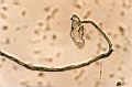 Cellulose making process of uracil base started at the level of molecule, micro fibril, misel and/or fibril, causes the changing of the relationship of the cells with all tissues, organs and systems and the ageing of the system, by escaping the control of the organism’s defence mechanism and by negatively affecting the functions of the cells related to the production of enzymes, hormones, secretes and neuro-secretions etc. Cellulose making process of uracil base started at the level of molecule, micro fibril, misel and/or fibril, causes the changing of the relationship of the cells with all tissues, organs and systems and the ageing of the system, by escaping the control of the organism’s defence mechanism and by negatively affecting the functions of the cells related to the production of enzymes, hormones, secretes and neuro-secretions etc.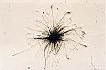 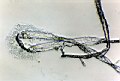 Urasil is related to many diseases such as BSE, Cancer and Alzheimer. Kat
|
|
|
|
Post by katinka on Mar 26, 2010 6:54:34 GMT -5
Cellulose making process of uracil base started at the level of molecule, micro fibril, misel and/or fibril, causes the changing of the relationship of the cells with all tissues, organs and systems and the ageing of the system, by escaping the control of the organism’s defence mechanism and by negatively affecting the functions of the cells related to the production of enzymes, hormones, secretes and neuro-secretions etc. Glutamate is the only amino acid that undergoes rapid oxidative deamination. This process leads to 2 toxic products: Hydrogen PeroxideAmmonia. Hydrogen Peroxide = Fenton's reaction.  Kammy and I were looking at Carnicom's last study where he mentioned this reaction to occur. A while back I was trying to 'tie' this all up: morgellons2.wordpress.com/2009/10/16/hormones-mineral-imbalance-susceptibility-for-morgellons-disease/fits... Kat |
|
|
|
Post by katinka on Mar 26, 2010 11:03:28 GMT -5
|
|
|
|
Post by skyship on Mar 26, 2010 11:55:51 GMT -5
Yes, when uracil was introduced into the genome, that began many diseases.
That was Kornberg and Nirenberg.
The triple in the downs syndrome is related to the addition of this. downs is related
to Alzheimers and the pancreas.
What we have now is a quadruplex.
A double strand inserted into the Watson Crick double strand helix.
Where the inorganic became part of the genome.
It fits........katinka
I believe Franklin was aware of this as well as Wilkins, but Crick and Watson
went with it.
Kerogens can be formed from this volcanic archaea and the nanobacterium.
Kerogens were the simulated PAHS
polycyclic aromatic hydrocarbons and am thinking this is the sphere formation.
Formed from a particle.
Space dust vs smart dust.
skyship
|
|
|
|
Post by katinka on Mar 26, 2010 18:57:01 GMT -5
Kerogens were the simulated PAHS polycyclic aromatic hydrocarbons and am thinking this is the sphere formation. Formed from a particle. Space dust vs smart dust. skyship Would fit, Sky, if you think about Nanobacterium found in Meteorites. Add environmental contaminates, water- and airpollution.....fossil fuel...Global warming and evtl. Cryptic pathogenisis = formation of PAHS. Fossil fuels are hydrocarbons, primarily coal and petroleum (liquid petroleum or natural gas), formed from the fossilized remains of dead plants and animals by exposure to heat and pressure in the Earth's crust over hundreds of millions of years.The burning of fossil fuels by humans is the largest source of emissions of carbon dioxide, which is one of the greenhouse gases that enhances radiative forcing and contributes to global warming. en.wikipedia.org/wiki/Polycyclic_aromatic_hydrocarbonen.wikipedia.org/wiki/FuelKat |
|
|
|
Post by skyship on Mar 27, 2010 2:04:09 GMT -5
Ah katinka, but this was created to look like PAHs from outer space., those are what Carl Sagan called "Tholins" form Titan, a Saturn moon and in interstellular space. Those are real, those are not simulated, from smart dust like kerogens are. To make it look like they came from space, when the real PAHS are in outer space. kerogens were used. from possibly Burgess Shale? and other places. www.spitzer.caltech.edu/features/articles/20050627.shtmlsimulated pahs from earth: Molecular simulation of polycyclic aromatic hydrocarbon sorption to black carbon Authors: J. J. H. Haftka a; J. R. Parsons a;H. A. J. Govers a Affiliation: a Earth Surface Sciences, Institute for Biodiversity and Ecosystem Dynamics, Universiteit van Amsterdam, 1018 WV Amsterdam, the Netherlands DOI: 10.1080/10629360902949336 Publication Frequency: 8 issues per year Published in: journal SAR and QSAR in Environmental Research, Volume 20, Issue 3 & 4 April 2009 , pages 221 - 240 Subjects: Applied & Industrial Chemistry; Chemistry; Environmental & Ecological Toxicology; Environmental Chemistry; Environmental Sciences; History & Philosophy of Mathematics; Simulation & Modeling; Abstract Strong sorption of hydrophobic organic contaminants to soot or black carbon (BC) is an important environmental process limiting the bioremediation potential of contaminated soils and sediments. Reliable methods to predict BC sorption coefficients for organic contaminants are therefore required. A computer simulation based on molecular mechanics using force field methods has been applied in this study to calculate BC sorption coefficients of polycyclic aromatic hydrocarbons (PAHs). The free energy difference between PAHs dissolved in water and in water containing a model structure of BC was calculated by thermodynamic integration of Monte Carlo simulated energies of transfer. The free energies were calculated with a hypothetical reference state that has equal free energies in both phases and is therefore cancelled in the calculated free energy difference. The calculated sorption coefficient of phenanthrene (log KBC = 5.17 ± 0.54 L/kg C), fluoranthene (6.33 ± 0.64 L/kg C) and benzo pyrene (7.38 ± 0.36 L/kg C) corresponded very well to experimental values available in the literature. Furthermore, an average spacing distance of 3.73 Aring between PAHs and BC was determined that is only slightly lower than an experimentally determined value of 4.1 Aring. The method applied in this study enables the calculation of the extent of PAH sorption to a soot surface for which no experimental values are available nor data for related compounds as required in quantitative structure-activity relationships.
Keywords: PAH; molecular simulation; Monte Carlo; free energy; statistical mechanics
www.informaworld.com/smpp/1728380859-97519432/content~content=a912561022&db=allskyship Polycyclic aromatic hydrocarbons. Actually it began in the Baltic Sea, or was it Sargasso Sea? A created phytoplankton was released into the fisheries. Because that was the protocol from the UN. Was it Rio or Toronto? mmmm not sure now. maybe euglena? mmmm not sure. or?  ?? www.biomareweb.org/downloads/ojaveer.pdfskyship |
|


 ...I recognized my typical way of writing..
